

Find it early, while it is small.
A low-dose breast X-ray that can pick up changes years before anyone could feel them. Screening and diagnostic mammograms, read by a radiologist.


Screening, or looking for an answer
Screening mammogram
For women with no symptoms at all. It looks for changes too small to feel, which is how most breast cancers found on mammography are found early enough to treat simply.
Diagnostic mammogram
For a lump, thickening, pain, skin dimpling, or nipple change or discharge. Extra views are taken and the radiologist tailors the study to what needs answering.
What the radiologist looks for
Calcifications
Tiny specks of calcium. Most are harmless, but certain shapes and clusters are worth following up.
Masses
A distinct shape with its own borders. Size, edges and density all change what it means.
Asymmetry
Tissue that shows on one side and not the other, or that is new since your last study.
Distortion
Tissue pulled out of its normal fan pattern without an obvious lump behind it.
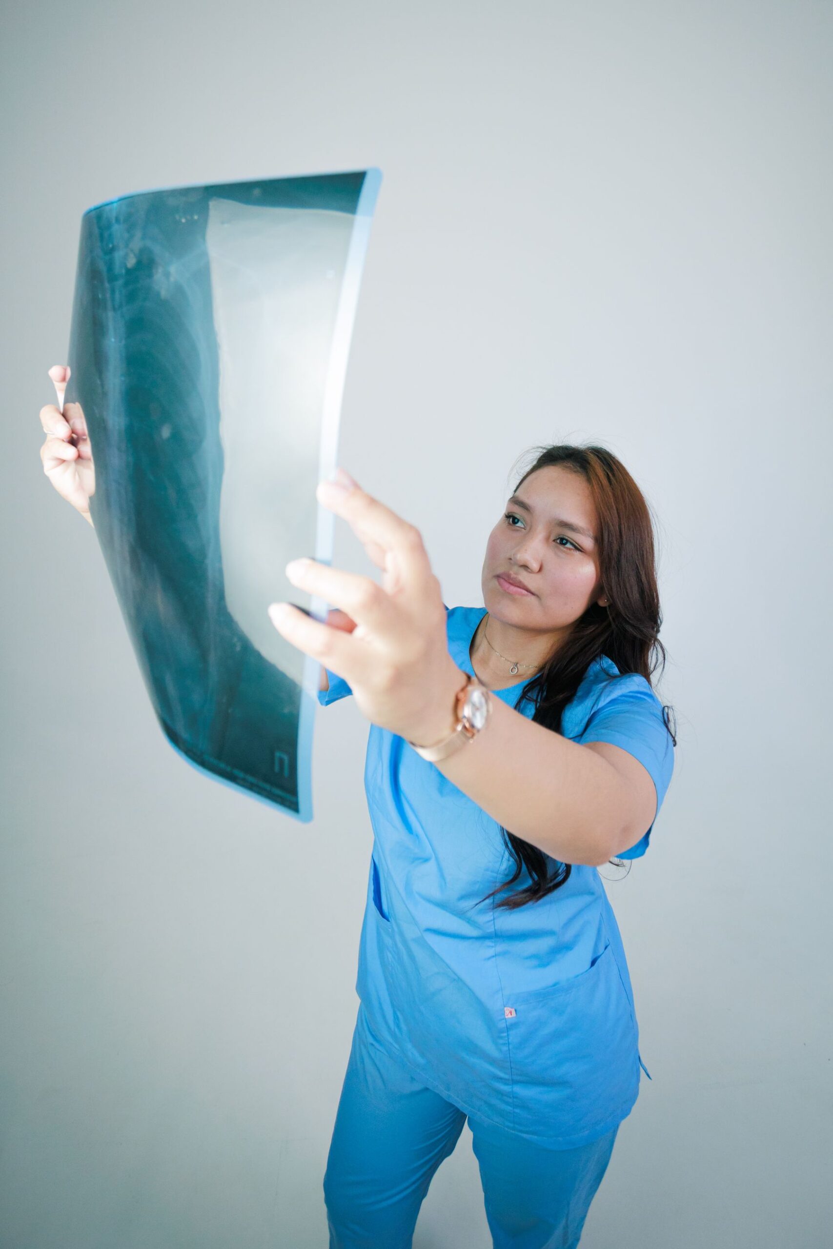

Read by a radiologist, next to your last films
A mammogram is most useful when it can be compared. Small changes between this year and last are often the finding that matters, which is why prior images are worth bringing.
The report goes to the physician who requested it, with a plain description of what was seen and what, if anything, should happen next.
What the appointment is like
Request
Bring the written request from your physician. Tell the desk if you have had a mammogram before and where it was done.
Positioning
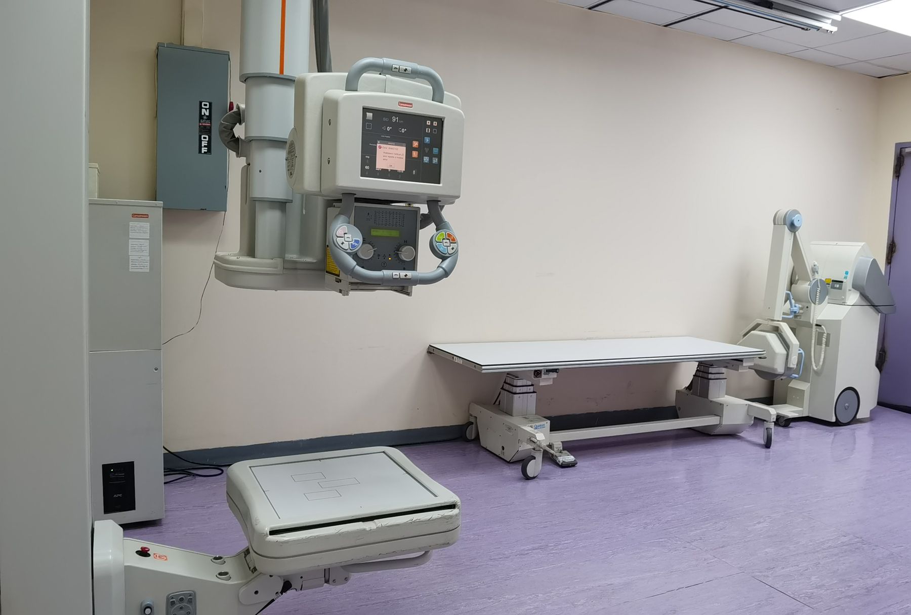
A radiologic technologist positions one breast at a time on the plate. Two standard views are taken of each side.
Compression
The plate presses firmly for a few seconds. It spreads the tissue so less radiation is needed and small details are not hidden.
Reading
The radiologist reads the images against any prior films and issues a report to the doctor who requested the study.
Before you come in
Book the week after your period. Breasts are least tender then, so the compression is easier to sit through.
Skip deodorant, powder, lotion and perfume on the day of the study. They can appear on the film as white specks that look like calcifications.
Wear a two-piece outfit. You will only need to undress from the waist up.
Bring prior mammograms or reports if you have them, even from another hospital. Comparison is half the value of the study.
Tell the technologist if you are pregnant or think you may be, if you are breastfeeding, or if you have implants.
Ready to book your mammogram?
Talk to the imaging desk about the study your physician asked for, or ask what a first screening involves.
Answers before your visit
Does a mammogram hurt?
There is firm pressure for a few seconds on each view. Most women describe it as uncomfortable rather than painful, and it passes quickly. Tell the technologist if it is too much, because positioning can be adjusted.
How much radiation is involved?
The dose is low, far below that of a CT scan. For women in the screening age range, the benefit of finding a cancer while it is small is generally considered to outweigh the small risk from the dose.
Do I need a request from a doctor?
Bring a written request if your physician has given you one. If you are arranging a screening mammogram yourself, the front desk will confirm what is needed before your appointment.
When will I get my results?
The radiologist report is released to the physician who requested the study. You can also collect a printed copy at the counter with your official receipt and a valid ID.
What if something is seen on the film?
Most findings are not cancer. You may be asked back for extra views or an ultrasound so the area can be looked at more closely. Your doctor will explain what the report means and what comes next.
Can I have a mammogram with implants?
Yes. Say so when you book, so the technologist can plan the additional views that are used to see around an implant.